ID Gallery: Blue Mold

View the 360 Rotator
showing multiple images of the external disease symptoms.

View the 360 Rotator
showing multiple images of the internal disease symptoms.
Blue Mold on Apples
Blue mold (primarily Penicillium expansum) is a very common postharvest fungal disease on apples worldwide. This disease is of economic concern to both the fresh-fruit industry and the fruit-processing industry because some strains produce the mycotoxin patulin, which can rise to unacceptable levels affecting the quality of apple juice.
For full description of Blue mold and management visit our Blue Mold Postharvest Disease page.
Click on images below to see full size.

Blue mold originating from infection of wound on fruit; decayed area brown, soft and watery, with a sharp margin; blue-green spore masses visible. Photo: CL Xiao, USDA-ARS.
Blue mold decayed tissue completely separable from the healthy tissue. Photo: CL Xiao, USDA-ARS.

Blue mold originating from infection of wound on a Granny Smith fruit; spore masses formed at the infection site. Photo: CL Xiao, USDA-ARS.

Calyx-end blue mold on a Fuji fruit; usually associated with drenched fruit. Photo: CL Xiao, USDA-ARS.

Blue mold on Fuji apple. Brown, soft and watery lesion with sharp margin. Originating from a wound. Notice white & green spore masses. Photo: A. Amir, WSU Plant Pathology.

Blue mold originating from wound infection on Granny Smith apple. Spore masses formed along the infection site. Photo: CL Xiao. USDA-ARS.

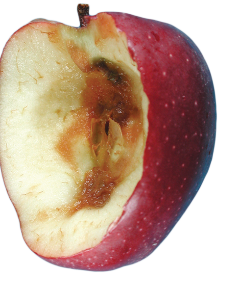
Cross section of blue mold lesion on Granny Smith apple. Sharp margin between decayed and healthy tissue. Photo: A. Amiri, WSU Plant Pathology.

Decayed tissue completely separated from the healthy tissue, leaving a “bowl-like” cavity. Photo: CL Xiao, USDA-ARS.

Advanced blue mold. Conidia are very light and easily dispersed to neighboring fruit with the slightest movement. Photo: A. Amiri, WSU Plant Pathology.

Honeycrisp apple showing the brown, soft, watery lesion with a definitive edge. Photo: TJ Mullinex, Good Fruit Grower.

Internal view of a Honeycrisp apple with blue mold infection. The decayed tissue was easily separated from the remaining healthy tissue. Photo: TJ Mullinex, Good Fruit Grower.

View looking straight into a Honeycrisp apple with blue mold infection originating from a side puncture. The decayed tissues was easily separable from the healthy flesh. Photo: TJ Mullinex, Good Fruit Grower.
Blue Mold on Pear
Click on images below to see full size.
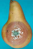
Blue mold on a Bosc fruit; watery decay lesion with white mycelium and blue spore masses. Photo: CL Xiao, USDA-ARS.
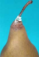
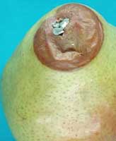
Stem-end blue mold on a Bosc pear fruit after an extended period of storage. Photo: CL Xiao, USDA-ARS.
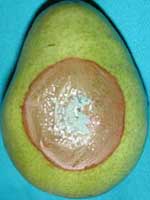
White mycelium and blue spore masses at the decayed area of a d’Anjou fruit. Photo: CL Xiao, USDA-ARS.
Stem-end blue mold commonly seen on d’Anjou pears, particularly after an extended period of storage. Photo: CL Xiao, USDA-ARS.