ID Gallery: Bull’s Eye Rot
Bull’s eye rot (BER) is a continuous threat to apple and pear storability in the PNW and in many other growing regions. Recent surveys from Washington State showed that BER accounted for 8 to 10% of overall decayed fruit and 40% of the surveyed orchards had BER at frequencies ranging between 1% and 75% (Amiri and Ali 2016). Some cultivars, such as Golden Delicious and Pinata are highly susceptible to Neofabraea spp., but other cultivars such as Fuji, Cripps Pink, and Granny Smith can also be at risk, especially when rainy conditions occur before harvest.
For more information about Bull’s eye rot and how to manage the disease see our page Postharvest Diseases: Bull’s Eye Rot.
Bull’s Eye Rot in Apple

Bull’s eye rot from a side wound infections. Photo: TJ Mullinex, Good Fruit Grower.

Bull’s eye rot lesion on the side of a Fuji apple. Photo: TJ Mullinex, Good Fruit Grower.

Bull’s eye rot on a Golden Delicious fruit. The lesion is flat to slightly sunken, brown to dark brown with lighter brown to tan center. Photo: CL Xiao, USDA-ARS.
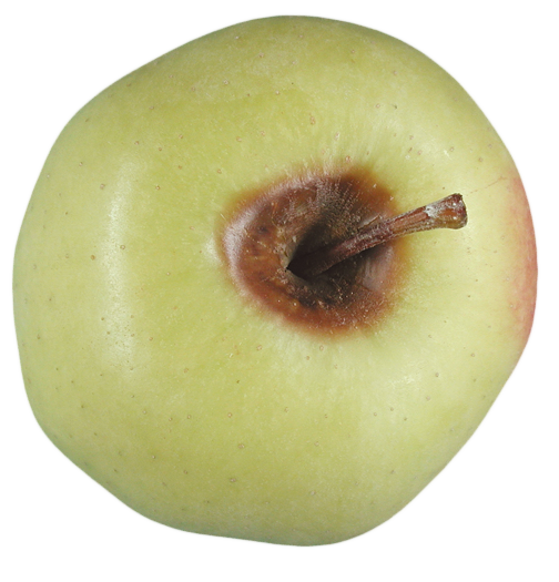
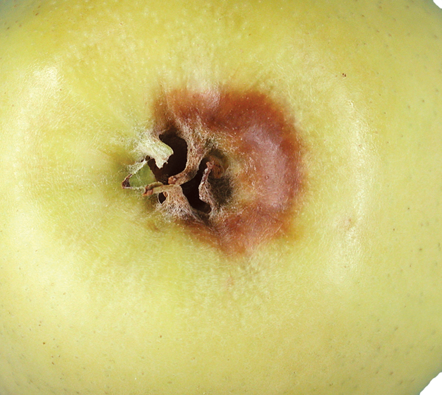
Bull’s eye rot originating from an infection in the stem bowl of this Golden Delicious apple. Photo: CL Xiao, USDA-ARS.
Bull’s eye rot lesion originating from an infection in the calyx area of this Golden Delicious apple. Photo: CL Xiao, USDA-ARS.

Bull’s eye rot originating from the stem bowl of this Gala apple. Photo: A. Amiri, WSU Plant Pathology.
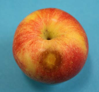
Bull’s eye rot originating from a lenticel infection on Gala. Compare to next image. Photo: A. Amiri, WSU Plant Pathology.

Bull’s eye rot originating from a lenticel infection on Gala. Note the difference with previous image. Photo: A. Amiri, WSU Plant Pathology.
Multiple Bull’s eye rot lesions on the side of a Golden Delicious apple. Photo: CL Xiao, USDA-ARS.
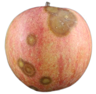
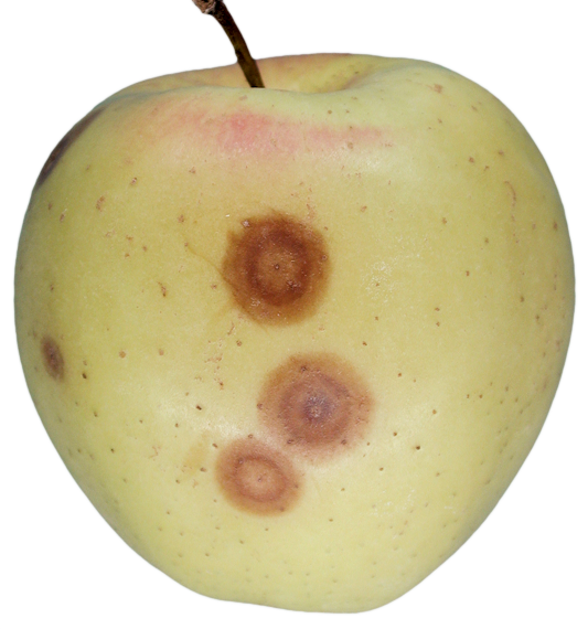
Multiple Bull’s eye rot lenticel lesions. Photo: Whitney Garten, WSU Horticulture Student.

Early Bull’s eye rot infection starting from a wound on the side. Note light brown in the center with a darker halo towards the outer edge. Photo: A. Amiri, WSU Plant Pathology.

A more advanced Bull’s eye rot infection originating from a side wound. Acervuli beginning to appear. Photo: A. Amiri, WSU Plant Pathology.
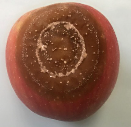
Advanced stage of Bull’s eye rot on Fuji apple. White mycelium and cream-colored spore masses clearly visible. Photo: A. Amiri, WSU Plant Pathology.
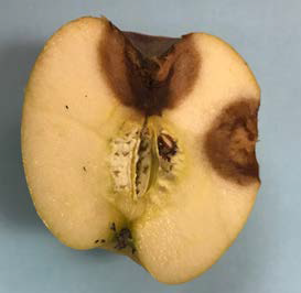
Cross-section of a Fuji apple showing internal symptoms of Bull’s eye rot originating from infections at both a lenticel and the stem-bowl. Note the sharp margin for each lesion. Photo: A. Amiri, WSU Plant Pathology.
Bull’s Eye Rot of Pear
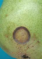
Bull’s eye rot on a d’Anjou pear fruit. Photo: CL Xiao, USDA-ARS.
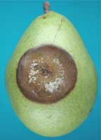
Advanced stage of Bull’s eye rot on a d’Anjou pear fruit; white mycelium and cream-colored spore masses present at the center. Photo: CL Xiao, USDA-ARS.

Advanced stage of Bull’s eye rot on d’Anjou pear. Cream-colored spore masses present at the center of the sunken lesion. Photo: A. Amiri, WSU Plant Pathology.