ID Gallery: Gray Mold

View the 360 Rotator
showing multiple images of the external disease symptoms.
Gray Mold on Apples
Gray mold (Botrytis cinerea) is a common postharvest disease on apples worldwide. This fungus has the ability to spread from decayed fruit to surrounding healthy fruit through fruit-to-fruit contact during storage. Because of this, significant losses as high as 20-60% are not uncommon after an extended period of storage, particularly on fruit that were not treated with fungicides prior to storage
For a full description of gray mold and it’s management visit our page Gray Mold Postharvest Disease.
Click on images below to see full size.

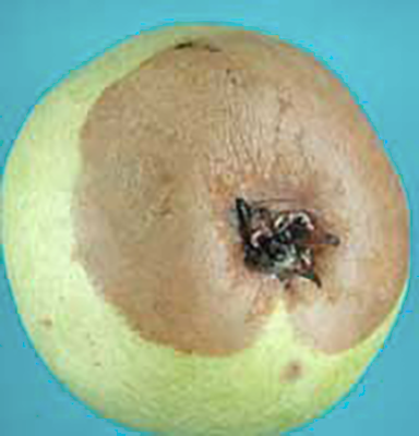
Gray mold originating from infection at stem bowl; gray spore masses may be visible at the diseased area under high humidity. Photo: CL Xiao, USDA-ARS.

Gray mold commonly originating from infection of wounds on the fruit; decayed area brown, spongy to firm; decayed tissue may become soft in very advanced stage. Photo: CL Xiao, USDA-ARS.

Gray mold originating from infection of the calyx of a Red Delicious; white to gray mycelium and gray spores may cover the decayed area under high humidity conditions. Photo: CL Xiao, USDA-ARS.

Gray mold at the stem-bowl of a Gala. Decayed area brown and spongy to firm. Photo: A. Amiri, WSU Plant Pathology.
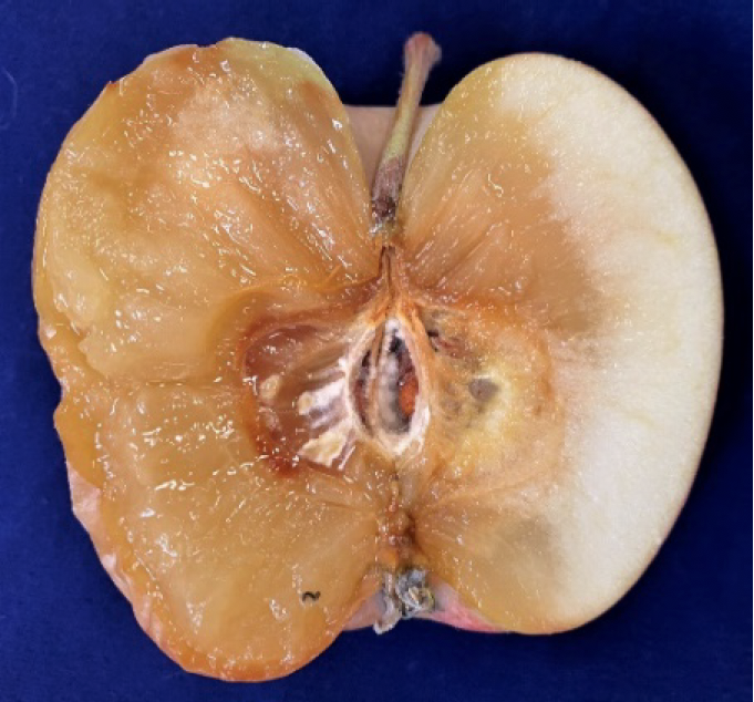
Internal view of a Fuji apple tissue infected by gray mold. Note the irregular margin and decayed tissue does not separate from healthy tissue. Photo: A. Amiri, WSU Plant Pathology.

Gray mold originating from side wound infection of a Granny Smith. Photo: A. Amiri, WSU Plant Pathology.
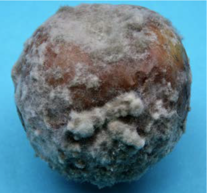
Advanced stage of gray mold. White to gray mycelium covering the decayed area under high humidity. Photo: A. Amiri, WSU Plant Pathology.
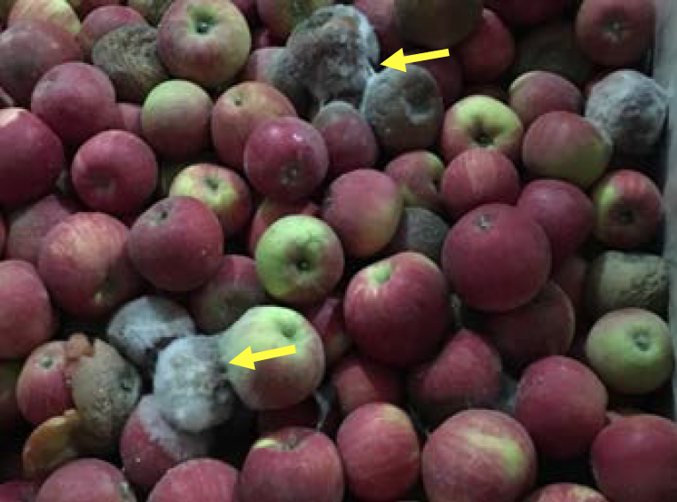
Nesting of gray mold due to fruit-to fruit spread on Honeycrisp in storage. Notice white to gray mycelium. Photo: A. Amiri, WSU Plant Pathology.

Gray mold infection at the stem end of a Honeycrisp apple showing fluffy white to gray mycelia. Photo: TJ Mullinex, Good Fruit Grower.
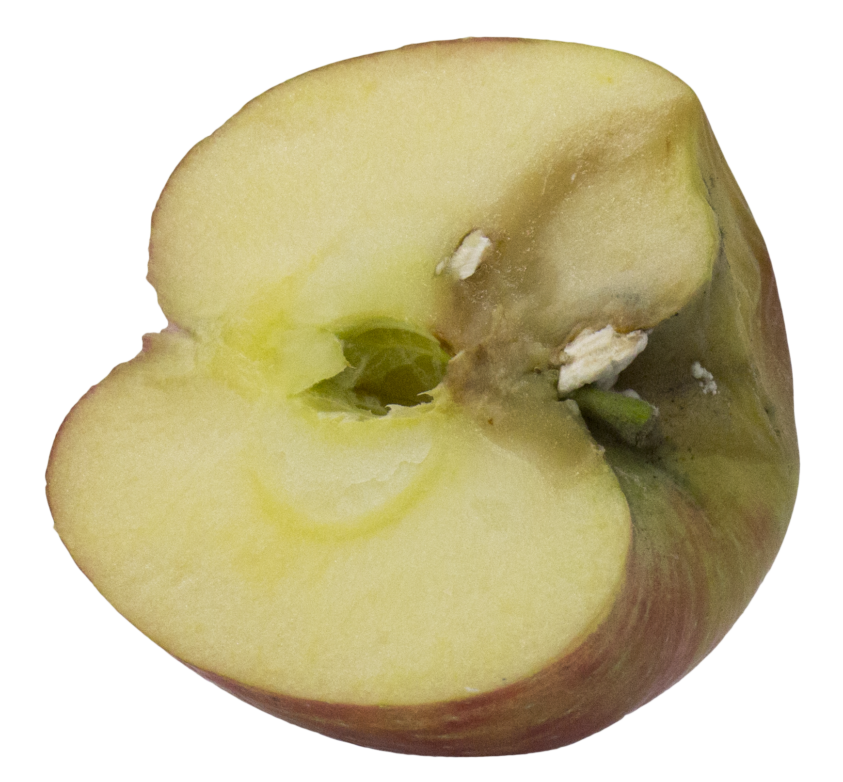
Interior view of gray mold on Honeycrisp originating from a stem bowl infection. Photo: TJ Mullinex, Good Fruit Grower.

External gray mold infection on Crips Pink apple not showing spores. Photo: TJ Mullinex, Good Fruit Grower.

Gray mold infection on Crips Pink apple with white mycelia and early spore formation in the stem bowl. Photo: TJ Mullinex, Good Fruit Grower.
Gray Mold on Pear
Click on images below to see full size.

Stem-end gray mold progressing toward the calyx-end commonly seen on d’Anjou pears. Photo: A. Amiri, WSU Plant Pathology.
Gray mold originating from calyx infection on a d‘Anjou pear. Photo: CL Xiao, USDA-ARS.

Gray mold originating from wound infection on a d’Anjou pear fruit. Photo: CL Xiao, USDA-ARS.
Sclerotia of B. cinerea formed on the surface of a decayed d ‘Anjou fruit at advanced stage. Photo: CL Xiao, USDA-ARS.